· News
The data is in: our biophoton microscope is ready for business
Martin Etzrodt, Open Science Institute
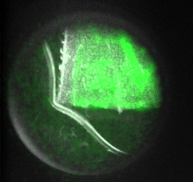
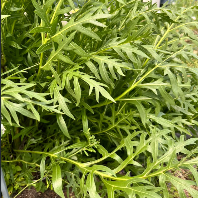
The data is in. On 8 and 9 June 2026, the biophoton microscope at openUC2 in Jena completed its first full measurement campaign. The method is simple to state: an injured leaf is given a small dose of hydrogen peroxide, placed in front of a camera cold enough and quiet enough to register single photons, and recorded for twenty minutes in the dark.
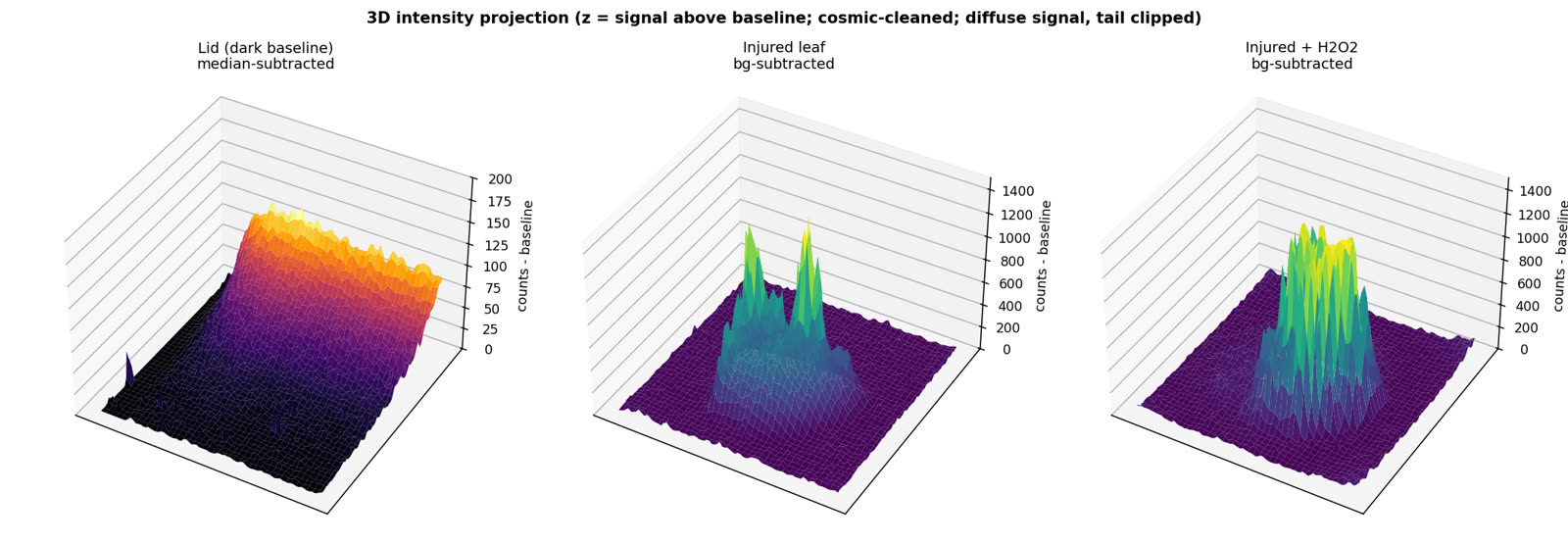
The result is clear. An injured compass plant (Silphium laciniatum) leaf treated with H2O2 emitted 1.56 times more total light than an untreated injured leaf, under identical camera settings. The brightest regions were roughly twice as bright, which indicates the additional light comes from localised reaction hotspots rather than a uniform rise across the leaf. Living tissue under stress emits light, and here that light was measured.
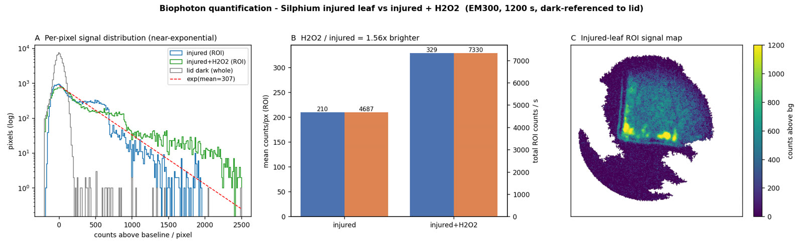
The character of the signal matters as much as its size. The per-pixel distribution is near-exponential, the signature of single photo-electrons amplified one at a time in the camera's electron-multiplying register. In other words, the detector was operating at its floor, counting light close to photon by photon. That is the regime required to measure emission this faint with confidence.
One limit should be stated plainly. Because the camera's true gain and sensitivity were not recorded in the acquisition metadata, the present numbers are relative rather than absolute. Closing that gap, with a calibration that converts counts into photons and carries a real error bar, is the next task, and it is being built directly into the open-source openUC2 control software so that a one-off correction becomes a tool the wider community can reuse.
The instrument
The measurements were run at openUC2 in Jena on the open-hardware microscopy platform developed by Benedict Diederich and his team, with the calibration work carried out together with Rainer Heintzmann and colleagues at the Leibniz IPHT in Jena. The rig itself is deliberately modest: a light-proofed enclosure, an Andor iXon EMCCD, a liquid cooler, and a laptop.
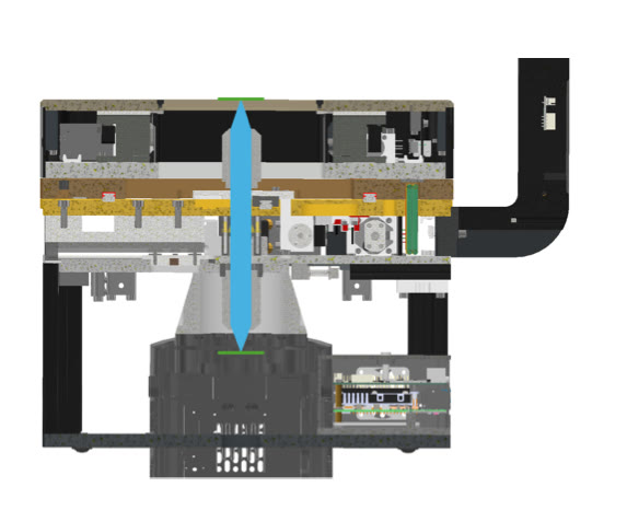
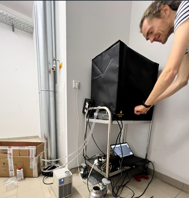
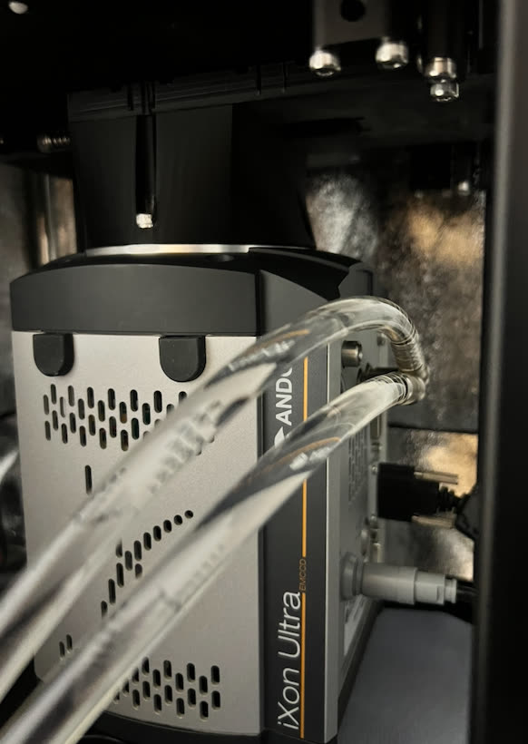
These are faint beginnings, in the most literal sense. The detector at the heart of the setup is a capable but costly scientific camera. Our aim is to develop a version of this instrument that performs the same measurement at a fraction of the price, and to bring it into as many hands as possible. An open instrument only advances open science if others can afford to build it.
Outlook
This campaign is the first step of a longer programme. The near-term work is to harden the Jena instrument into a fully reproducible rig, complete the calibration, and widen the study across plant species and stress conditions with proper biological replicates. From there, the aim is to use these plant screens to connect specific photon signatures to the underlying biology, and, further out, to translate the deep-cooled bench sensor into an affordable and eventually wearable detector, carrying the same measurement from a leaf on a bench toward the human body.
The faint light that living things emit was a recurring subject at our first community gathering at Etherlaken in June. Now it is something we can measure.
Licensed under CC BY 4.0. Free to share and adapt with attribution. (link opens in a new tab)